Horse Leg Bones Diagram - The Horse Veterian Key : Abnormal or crooked front legs can lead to lameness by putting stress on the following.. They are joined to the spine through the sacroileac joints and allow transfer of propulsion to the hind legs. The tendons of the lower leg are the deep digital flexor tendon, superficial digital flexor tendon, lateral digital flexor tendon and common digital flexor tendon. Within 20 minutes of birth a foal may stand, and within hours can be ready to run at speeds no human athlete will ever achieve. Horse anatomy diagrams legs via. Horse stifle joint anatomy via.
This system works together to support horses weight when it stands up also works to diminish compression during movement which helps to horse to avoid injury to their limbs. Horse stifle joint anatomy via. Horse vs human heather smith thomas made a beautiful comparison, in her book the horse conformation handbook, between the anatomy of the horse's lower leg and that of the human hand. » science abc from www.scienceabc.com 13.05.2021 · horse leg bone diagram : The healing process below the knee or hock is slow due to the lack of blood supply to the tissue and bone (there is no muscle on the lower legs of a horse).
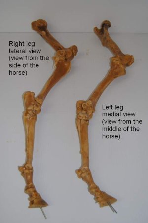
Bones Of The Hind Leg Part One from www.rodnikkel.com Which is the oldest horse on the diagram? Talking about horse anatomy worksheets printable, we have collected some related images to complete your references. Most lameness occurs in the forelimbs because 65% — 70% of the horses weight is carried by the front legs. The horses legs and hooves are also unique, interesting structures. There are many possible diagrams of the anatomy of horse tissues. Hoof and lower leg structure. The eohippus horses part b. The photograph shows the laminae which keep the hoof wall tightly bonded to the internal structures.
Bones are connected to muscles via tendons and other bones via ligaments.
Horse leg structure there are also many fossil remains of horse leg bones. This line should evenly split the forearm, knee, cannon, fetlock, pastern and hoof. And 2 years, but the rest of the bone does not stop growing until the horse is 5 or more. In our website, we are persons who very treasure creativity from every one, with no exception. The top part of the hind limbs consists of three fused bones, called the ileum, ischium, and pubis. Bones are connected to muscles via tendons and other bones via ligaments. Cartilage extends backwards and upwards from this bone. Abnormal or crooked front legs can lead to lameness by putting stress on the following. A thin band of white above a horse's hoof. The power propulsion system and major defensive tool, a horse's rear. Hoof and lower leg structure. Sock (sometimes called an anklet): Horse body parts diagram, horse skeleton diagram and animal nervous system diagram are some main things we want to present to you based on the gallery title.
Abnormal or crooked front legs can lead to lameness by putting stress on the following. This system works together to support horses weight when it stands up also works to diminish compression during movement which helps to horse to avoid injury to their limbs. How many front toes did the oldest horse have? The legs of a horse are made up of a system of muscles, tendons, ligaments, and connective tissue. The photograph shows the laminae which keep the hoof wall tightly bonded to the internal structures.
Chestnut Horse Anatomy Wikipedia from upload.wikimedia.org At this stage of life, even with this exceptionally early development, horses have only 17% of their mature bone mineral content. Abnormal or crooked front legs can lead to lameness by putting stress on the following. This is a free printable worksheet in pdf format and holds a printable version of the quiz horse leg bones. The horse's back leg is homologous to a human's leg, too, but we are going to focus on the foreleg and arm as our example for this blog.) beneath (or outward from) the humerus there are two bones, side by side: Diagram of leg bones and joints via. Proper hoof care, good exercise management. It is made up of the ilium, the ischium, and the pubis.at the junction of these three bones is a cavity called the acetabulum, which acts as the socket of the hip joint.the pelvic cavity is larger in diameter in the mare than in the stallion, providing more room for the foal during birth. Lower extremity arterial duplex via.
The bones of the horse skeleton are held together with ligaments, tendons and muscles.
The hoof is heavily supplied with blood through the two arteries which run down the back of the leg and into the foot. One bone works in relation to another. Made up of the os coxae, the largest of the flat bones in a horse. The limbs of the horse are structures made of many bones, joints, muscles, tendons and ligaments that support the weight of the horse's body. There are many possible diagrams of the anatomy of horse tissues. From the front of the horse, you should be able to draw a straight line from the point of the shoulder down the center of the leg. For example, the body part that is called a horse's knee is actually made up of the carpal bones that correspond to the human wrist. Talking about horse anatomy worksheets printable, we have collected some related images to complete your references. Horse anatomy diagrams legs via. The eohippus horses part b. The legs of a horse are made up of a system of muscles, tendons, ligaments, and connective tissue. The limbs play a major role in the movement of the horse, with the legs performing the functions of absorbing impact, bearing weight and providing thrust. Within 20 minutes of birth a foal may stand, and within hours can be ready to run at speeds no human athlete will ever achieve.
This diagram shows the superficial layer of the tissue. Proper hoof care, good exercise management. For example, the body part that is called a horses 'knee' is actually the carpal bones that correspond to the human wrist. From the front of the horse, you should be able to draw a straight line from the point of the shoulder down the center of the leg. This is because there are many layers of muscles.
Understanding Your Horse S Hock Health Dressage Today from dressagetoday.com The bones of the horse skeleton are held together with ligaments, tendons and muscles. And 2 years, but the rest of the bone does not stop growing until the horse is 5 or more. In this picture it shows the muscles that are closest to the surface of the skin, making them superficial. The power propulsion system and major defensive tool, a horse's rear. How many front toes did the oldest horse have? The tendons of the lower leg are the deep digital flexor tendon, superficial digital flexor tendon, lateral digital flexor tendon and common digital flexor tendon. This diagram shows the superficial layer of the tissue. Lameness problems from the fetlock down are usually caused by excessive.
They are joined to the spine through the sacroileac joints and allow transfer of propulsion to the hind legs.
Horse vs human heather smith thomas made a beautiful comparison, in her book the horse conformation handbook, between the anatomy of the horse's lower leg and that of the human hand. The pedal bone itself has an unusually high density of blood vessels within it. It also includes the joints of the hip, stifle, hock, fetlock, pastern, and coffin. The limbs play a major role in the movement of the horse, with the legs performing the functions of absorbing impact, bearing weight and providing thrust. The hoof is heavily supplied with blood through the two arteries which run down the back of the leg and into the foot. Both pelvic and thoracic limbs contain the same number of bones, 20 bones per limb. If the angle at witch these bones are working is compromised, the joint becomes unevenly stressed and injury to the tendons and ligaments. Sock (sometimes called an anklet): Principles of bone development in horses. Includes the pastern but doesn't cover the fetlock. The line leading from eohippus to the modern horse exhibits the following evolutionary trends: They are joined to the spine through the sacroileac joints and allow transfer of propulsion to the hind legs. The power propulsion system and major defensive tool, a horse's rear.
Their leg bones are proportioned differently from those of a human leg bones diagram. They are joined to the spine through the sacroileac joints and allow transfer of propulsion to the hind legs.


0 Komentar